


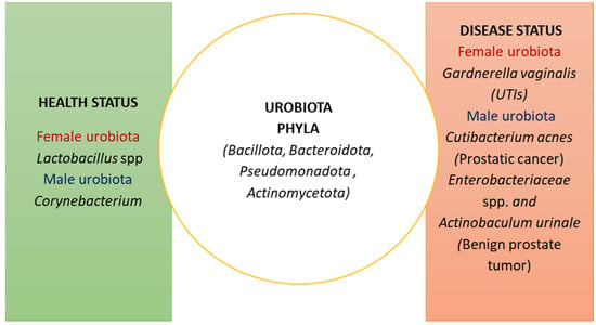
Pathophysiology 2024, 31(2), 298-308; https://doi.org/10.3390/pathophysiology31020022 - 8 Jun 2024
Abstract
►
Show Figures
Vaginal agenesis (VA) is frequently associated with mullerian agenesis. VA treatments include mechanical dilation and surgical vaginoplasty. We created a vaginal expansion sleeve (VES) as a novel device to progressively lengthen the vaginal canal. This study evaluated the histologic effects of the VES
[...] Read more.
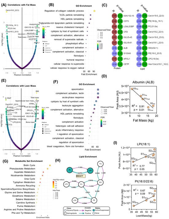
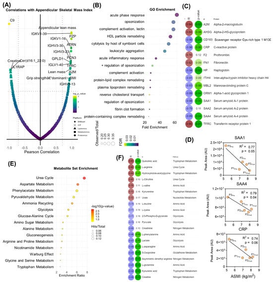
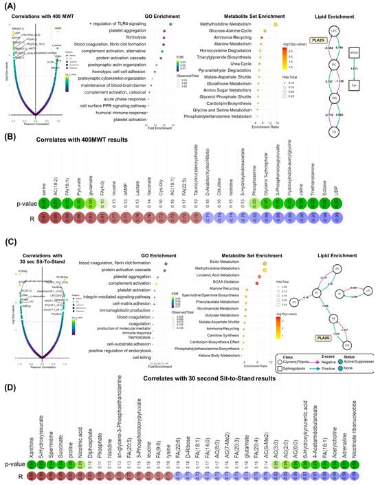
Vaginal agenesis (VA) is frequently associated with mullerian agenesis. VA treatments include mechanical dilation and surgical vaginoplasty. We created a vaginal expansion sleeve (VES) as a novel device to progressively lengthen the vaginal canal. This study evaluated the histologic effects of the VES on rat vaginal tissue. The VES is a spring-like device made of proprietary woven cylindrical material and flat resin caps. The VESs were constructed as 25–30 mm, pre-contracted springs, which were secured into the vaginas of six Sprague Dawley rats and allowed to re-expand post-surgically. After one week, the VESs were removed, and the vaginas were harvested and measured in length. Test (n = 6) and control (n = 4) formalin-fixed paraffin-embedded tissues were stained with hematoxylin and eosin (H&E), Masson’s trichrome, and anti-Desmin antibodies. The VESs achieved significant vaginal lengthening. The mean vaginal canal length increased from 20.0 ± 2.4 mm to 23.8 ± 1.2 mm after removal of the VESs (n = 6, p < 0.001), a 19% increase. There was a positive correlation between the expander/tension generated in the vagina and the amount of acute and chronic inflammation. H&E staining revealed increased submucosal eosinophilia in five of the six test tissues. One VES sample that was lengthened to 30 mm long showed evidence of lymphocytic and neutrophilic inflammation. Desmin immunostaining and Masson’s trichrome stain revealed a thinner muscularis with more infiltrative fibrous tissue between muscle fibers in the test tissue compared to the control tissue. Although effective, the VES may provoke at least a transient increase in eosinophils consistent with a localized immune reaction during muscularis remodeling.
Full article

Figure 1


